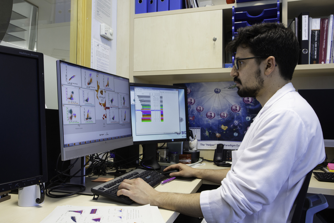
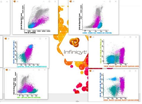
The World Health Organisation (WHO) classification of tumours proposes a multi-parametric approach to disease diagnosis, integrating clinical, morphological, phenotypic and genotypic data characteristic of each entity, including immunophenotypic study by flow cytometry. This is based on the use of laser light beams that intercept cells as they pass through the flow chamber of the cytometer. The cells are sorted according to their size, complexity and the various markers used in the analysis, usually in the form of specific antibodies labelled with different fluorochromes. The presence or absence of expression of these markers is measured simultaneously as a relative percentage and, in some cases, as absolute values of each specific cell type. New generation multiparametric flow cytometry (NGF) provides highly sensitive and accurate cell analysis for the separation, sorting and quantification of cells in suspension by acquiring millions of cells using protocols standardised by professional societies or consortia.

At Catlab we work with a BD FACSLyricTM digital cytometer, consisting of 3 lasers with a configuration of 12 fluorescence channels and 14 parameters with a maximum acquisition rate of 35,000 ‘events’ per second, and a BD FACSCantoTM II cytometer, also digital, with 3 lasers, a configuration of 8 colours and 10 parameters.

In parallel, we use the InfinicytTM analysis software, a tool patented by the Euroflow Consistory, which allows us to perform a multi-parametric analysis of millions of complex data contained in the files generated by the cytometers, using common markers in the different staining panels of the cells of cellular interest studied. This cell analysis mechanism allows for improved diagnosis, treatment and monitoring of leukaemias, lymphomas and myelomas, as well as other pathologies.
The Cytometry Area provides services to different medical specialties, mainly Haematology, Pathological Anatomy, Digestology, Neurology, Internal Medicine, Rheumatology, Ophthalmology and Paediatrics. We work with different types of samples: peripheral blood, bone marrow, tissues, adenopathies and fluids.
The studies performed in daily clinical practice can be classified into oncohaematological diagnostic studies and immunological diagnostic studies:

Oncohematologic Diagnosis

  • Screening for Chronic Lymphoproliferation Syndrome.
  • Chronic Lymphoproliferation B (SLPC-B) Syndrome: LLC, LNH Follicular, Mantell´s, Tricoleukemia,...
  • Chronic Lymphoproliferation Syndrome T/NK: LGL, LLC-T, Sézary, ..
  • Quantification of the Protein ZAP-70 as a predictive factor in LLC-B.
  • Immunophenotype of Acute Leukemia: LAM, LAL-B, LAL-T,...
  • Immunophenotype of plasma cells: MM, MGUS, Plasma cell Leukemia.
  • Quantification of Hematopoietic Progenitors “Stem Cells CD34+” with a single platform (expressed in percentages and in absolute values) in samples of peripheral blood (pre-apheresis)for the autologous transplant of peripheral blood (TASP).
  • Immunophenotype of bone marrow in Myelodisplasic and Myeloproliferative Syndromes

Immunologic Diagnosis

  • Quantification of Lymphocyte subpopulations T, B and NK with a single platform (percentage and absolute values).
  • LymphocyteT subpopulations in HIV.
  • Lymphocyte subpopulations in BAL.
  • Determination of the Antigen HLA-B27 (Associated with Andylosing Spondylitis and other rheumatologic diseases).
  • Immunophenotype of Duodenal Intraepithelial Lymphocytes to discount refractive celiac disease.

Citometry Images